


It is very common for children to fall down and sustain lacerations (medical term for cuts) over the face and limbs.
Although most wounds can heal with wound care and time, the aim of suturing a laceration is to oppose the edges as perfectly as possible. This is especially needed if the cut is deeper and underlying fat is exposed. A scar is inevitable — thus the aim is to make it as thin and inconspicuous as possible.
Plastic surgeons have unique knowledge of facial anatomy and are challenged every day to make scars beautiful. Using microsurgical suturing technique and magnifying loupes, the best possible result can be achieved.
Toilet and suture (T&S) can be performed in the Emergency Department under local anaesthesia. A gentle and patient approach will minimise trauma for the young patient. For larger and deeper cuts, this can be done under sedation or general anaesthesia in the operating theatre.
The procedure may typically be covered under accident insurance or hospitalisation insurance, depending on the terms of one’s insurance policy.
Although absorbable sutures can be used for the external layer, using non-absorbable sutures reduces the scar formation and fibrosis that arises from absorption of the suture.
After sutures are removed at 5-7 days, the scar will initially appear raised and pinkish for the first 3 months. The scar tissue will be remodelled from the third month onwards, resulting in scar flattening and improvement.
Scar management with sunblock, silicone scar cream, massage, injections and lasers will optimise the result.
Plastic surgeons are often involved in the assessment and treatment of skin lesions and subcutaneous lumps. Although the majority are benign (non-cancerous), early evaluation is vital if there are suspicious symptoms or signs.
These include large size or increase in size over a few months, irregular appearance (shape or colour), pain or bleeding. If it has not changed at all in years, it is usually benign.
If in doubt, it is better to seek the advice of a medical professional. A biopsy may sometimes be required to ascertain the diagnosis.
As an alternative to excision biopsy (complete removal), superficial skin lesions such as benign moles can be vaporized using a carbon dioxide laser. The small crater that forms heals over a few days, forming a flat pink spot.
Sun avoidance, sunblock and sometimes anti-pigmentation creams are critical to avoid post-inflammatory hyperpigmentation, when the spot can turn dark brown due to increased melanin activity.
Depending on the depth of the mole, a small raised scar can result if the deeper dermis was entered, although the scar typically flattens with time.
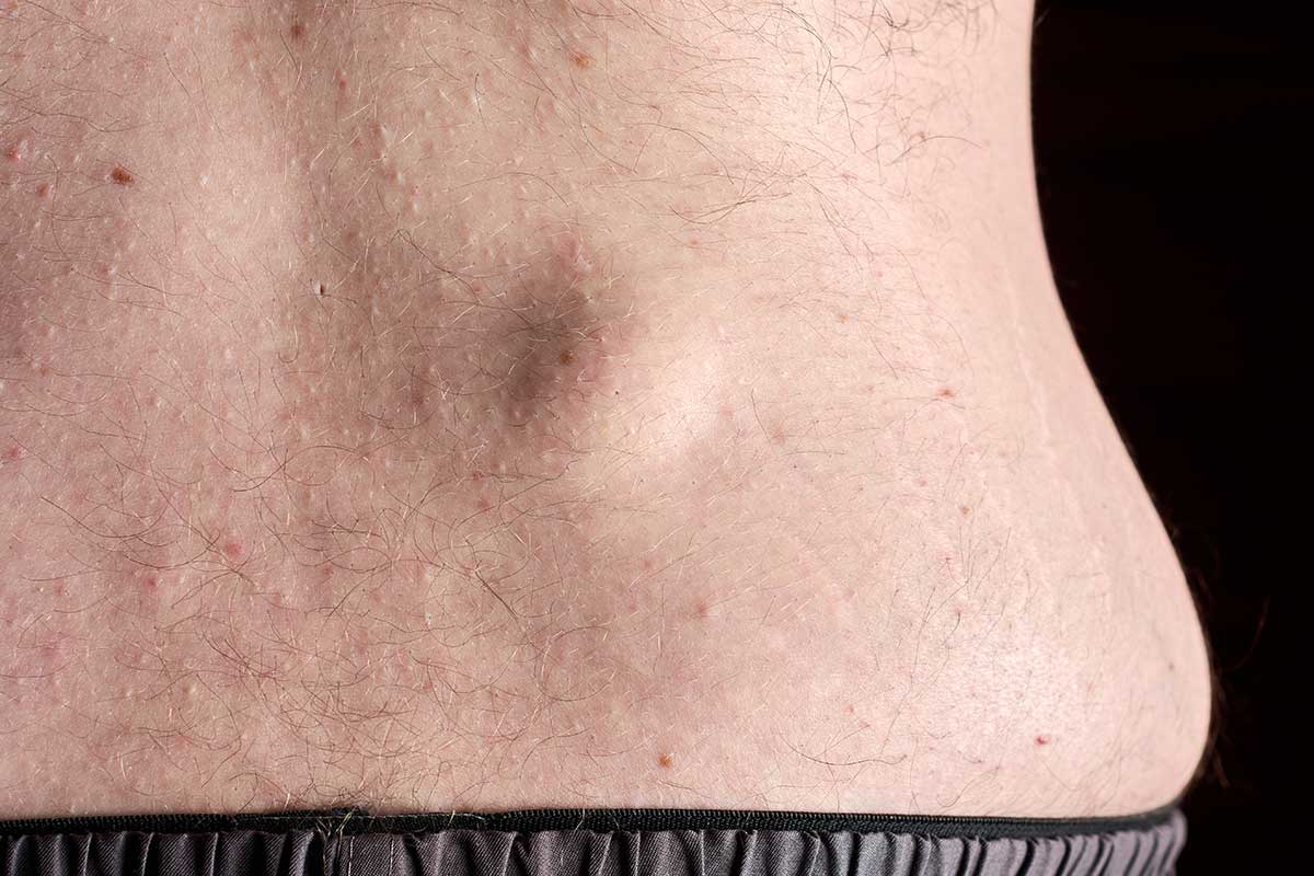
The most common subcutaneous lumps are lipomas or epidermoid cysts.
A lipoma is a non-cancerous fatty tissue growth under the skin. It should be removed if it is very big, growing, painful or if it bothers the patient. An epidermoid cyst is another non-cancerous nodule filled with keratin material.
As these lumps can occur over the face or noticeable areas, it is important to make the incision as small as possible. The resultant scar can also be reduced with gentle, meticulous suturing technique and early post-operative scar management.
Conversely, a lump can be suspicious if there are signs such as an irregular edge, fixation to deeper tissue or growth over time. Although less common, these should be evaluated as early as possible.
If a skin cancer is detected, wider removal with clear margins is required. This is because surgery offers the best chance of “clearing” the tumour and lowers the risk of it coming back.
The resulting defect (or hole) tends to be bigger and closing the wound directly may not be possible. Surrounding structures such as the eyelids or nose may be distorted by too much tension. A flap of surrounding tissue is then designed and “turned in” to close the hole, in what is termed “local flap closure”.
Using lasers and injections, the area can be blended in with the surrounding skin. Further minor revisions are sometimes needed to make the results even better.
Some patients wear their scars as a badge of honour, though many more are troubled by problematic scars. Certain scars are in more exposed areas that cannot be covered by clothing, such as the face, neck and hands. Less than ideal scars may be broad and depressed, thick and discoloured. There may also be “dog ears” – excess tissue at the ends of scars.
Due to factors such as genetics and scar location, some scars can be more raised than normal. Such hypertrophic scars do not extend beyond the boundaries of the original scar and usually become flatter with time. Keloid scars, on the other hand, usually grow and expand beyond the original scar. They also cause itch and pain.
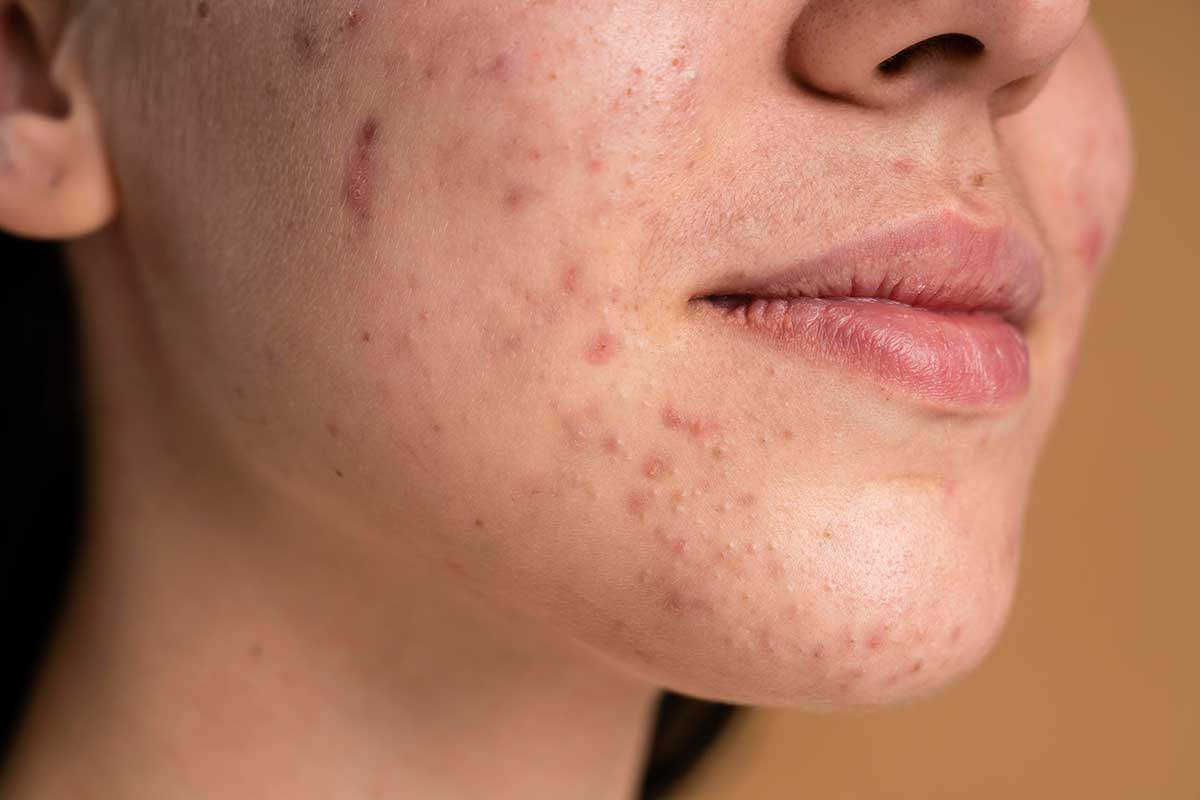
Basic scar treatment consists of silicone-based gel or tape, sunblock and massage. This is generally sufficient for normal and hypertrophic scars. Certain types of acne scars are treated by subcision and filler or fat injection.
A keloid scar requires much more intense and frequent treatment to control its growth. This consists of steroid tape, injections and laser treatment. In some cases, excision and local flap treatment or even radiation therapy is required.
Our plastic surgeons regularly managed burn patients at the Burn Centre in Singapore General Hospital. The Burn Centre is the only specialised facility and tertiary referral centre managing major burn injuries in Southeast Asia. Our surgeons are thus well-versed in the treatment of all types of burn wounds.
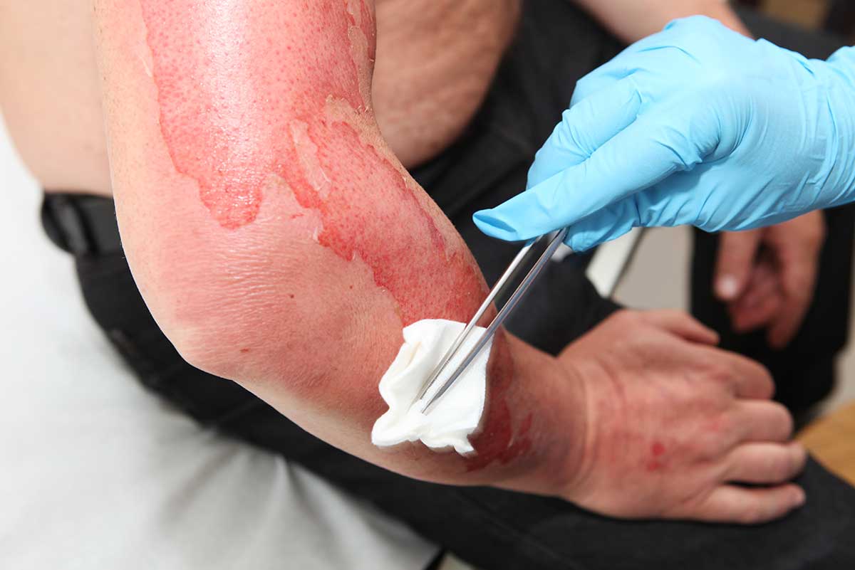
Scalds and burns are common injuries at home or in the workplace. Although a burn wound often starts out superficial, due to the process of burn conversion, the burn wound becomes deep. A deep wound can get infected easily. Even if heals, the prolonged healing time (>21 days) stimulates the development of thick, problematic scars.
It is important to seek consultation early as there are new biological dressings (such as Biobrane) that can reduce the conversion risk. They enable the wound to heal on its own with minimal scarring. Specialized wound care can hasten healing.
A burn wound that is deeper will require excision and skin grafting. Post-operative care consists of compression dressings, silicone sheets, lasers and injections.
An acute wound heals within 3 weeks. If healing is prolonged beyond this, the wound has become sub-acute (3 weeks to 3 months) or chronic (more than 3 months).
For chronic wounds, there is usually an underlying pathology that produces a delay in the healing process. This includes peripheral vascular disease, diabetes, infection and so on. These factors should be optimized to create a better wound healing environment.
Cleaning the wound and using advanced wound products such as vacuum-assisted therapy can sometimes be sufficient to kickstart the healing process again.
Procedures such as skin-grafting, dermal matrix application or flap closure can help expedite the process when the wound bed is ready.
Fractures of the facial bones, such as nasal bones, cheekbones and jaw bones can result in significant impact on facial appearance and function. Common causes include falls, blunt trauma and road traffic accidents. A CT scan is required to evaluate facial fractures accurately.
The blood supply to the face is excellent, as a result, facial bone fractures heal within 3-4 weeks. Our priority is to put back the fracture fragments in as anatomic a position as possible. The bony position is then stabilized with a few mini-titanium plates. All this is accomplished with hidden incisions such as within the mouth.
When critical structures such as facial nerves or vessels are interrupted, they can also be repaired under an operating microscope.
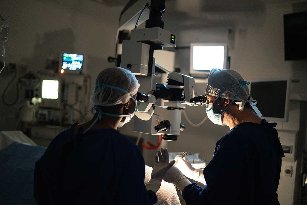
As reconstructive microsurgeons, we specialize in defects (tissue loss) of all sizes and complexity spanning the whole face and body. These defects occur after cancer removal or accidents. Beyond filling a “hole” with tissue, advances in the last few decades enable us to tailor a like-for-like reconstruction. Depending on what needs to be restored, soft tissue flaps comprising different vascularized components such as nerves, fascia, muscles can be harvested while minimizing impact on the donor site. The flap is transplanted to where it is required. The blood vessels are then connected to those in the recipient site using the operating microscope, with sutures finer than a human hair (microvascular anastomosis).
Once integrated with the body by 1-2 weeks, this internal tissue transplant is stable and functions like any other body part. Using the concept of the reconstructive elevator, microsurgical tissue transfer to cover defects is very common nowadays. This is because it offers an abundance of healthy tissue for a durable and aesthetic reconstruction.
Despite such sophisticated methods, there are still many instances where simpler loco-regional flaps, skin grafting and dermal matrices are the preferred method. A case-by-case evaluation and discussion with the patient are thus required.

The lymphatic system consists of multiple lymphatic channels that drain into lymph nodes. Lymph nodes are bean-shaped glands that monitor and cleanse the lymph as it filters through them. Lymph nodes may need to be removed as part of cancer surgery —for example, axillary (armpit) lymph nodes for breast cancer treatment.
When this happens, the lymphatic flow is blocked and the protein-rich fluid accumulates in the arms or legs. This is termed lymphedema. In some patients, the swelling is well-controlled by compression garments and they have a stable course without worsening.
In others, the main lymphatic channels become progressively scarred, resulting in backflow of fluid into the skin. The skin then becomes thickened and loses pliability. Repeated skin infections (cellulitis) can result due to impairment of immune defences in the abnormal skin.
The lymphatic fluid is also rich in lipids (e.g. cholesterol and fatty lipids). Besides fluid and fibrotic tissue, fat also accumulates over time. This contributes to the increase in weight and a feeling of heaviness. Severe cases can affect the ability to move the affected limb.
It is essential to follow-up with a physiotherapist specializing in lymphedema. The aim of conservative therapy is simply to manually push the lymphatic fluid upwards, back towards the heart. Treatment consists of lifelong compression bandages and stockings, massage and skin care.
Compression gloves and stockings are custom made after taking accurate measurements of the patient’s limb. They have to be worn every day, except when bathing or sleeping. They also have to be replaced or tightened every 6-12 months when they become loose, as a certain critical pressure is needed.
The affected arm or leg also needs to be elevated on a pillow at night.
New advances in the last 20 years have focused on “physiologic methods”. These aim to re-establish new drainage pathways for lymphatic fluid to flow.
For example, lymphatic channels can be joined to superficial veins (Lymphatic-venous bypass). This is a minimally-invasive procedure as the lymphatics and veins are just under the skin and can be accessed by targeted incisions. This is suitable for earlier stage patients in whom lymphatic channels are still present.
In later stages, a more viable option would be to transplant lymph nodes or lymphatic channels from elsewhere. In successful cases, patients can be less reliant on compression stockings and also experience resolution of repeated infections.
Some of the swollen tissue can be removed with liposuction or open excision. This reduces the weight of the limb and can improve mobility. However, as these are not physiologic procedures, it is essential to continue complex decongestive therapy as usual.